Methods
- Basic Light Microscopy
-
Morphology on millimeter and micrometer scale in reflection and transmission of bulk material and thin film.
- Confocal Microscopy
-
High-resolution imaging (of biological specimens, providing detailed, sharp images, Depth selectivity (allowing for the imaging of different layers within a specimen), Three-dimensional imaging (of specimen by compiling a series of two-dimensional images at different depths), Live cell imaging (allowing researchers to observe cellular processes in real time), Multi-color imaging (enabling the visualization of multiple targets within a single specimen), Reduced background noise and increased contrast (by using a pinhole to exclude out-of-focus light), Photobleaching and phototoxicity reduction (by only illuminating the plane of focus), Co-localization studies (to determine if two or more proteins are located in the same area of a cell), Fluorescence recovery after photobleaching (FRAP, which allows for the measurement of molecular mobility.
- Fluorescence Microscopy
-

Particle identification by autofluorescence and after staining with fluorescence dyes on filters in environmental, biological , medical and food samples, mostly as correlative spectroscopy in combination with optical and FTIR- and/or Raman microscopy, see cluster Spectroscopy.
- Raman Microscopy /FTIR Microscopy
-

see cluster Spectroscopy, mostly as correlative spectroscopy in combination with optical and/or fluorescence microsspectroscopy
- Scanning Electron Microscopy (SEM)
-
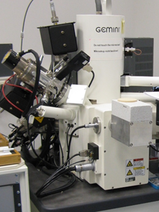
Morphology on micrometer and nanometer scale of bulk material, thin films and nanoparticles. Cross-sections by focused ion beam (FIB). Elemental mapping by energy dispersive X-ray spectroscopy (EDX). Variable-pressure SEM for imaging of humid specimens.
- Transmission Electron Microscopy (TEM)
-

Morphology and structure on nanometer scale of thin films and nanoparticles (bulk not available any more). Cryogenic TEM (cryo-TEM) of nanoparticles in aqueous dispersions. Elemental mapping by energy filtered imaging (EFTEM) and electron energy loss spectroscopy (EELS).
- Scanning Force Microscopy (SFM)
-
See Atomic Force Microscopy.
- Atomic Force Microscopy (AFM)
-
Topography of surfaces on nanometer scale, spatially resolved measurements of mechanical parameters (Young’s modulus, adhesion, energy dissipation), distribution of electric charges or magnetic domains, surface potential, electric conductivity, melting and solidification behavior, chemical composition.
- Atomic Force Microscopy (AFM-IR)
-
See cluster Spectroscopy
- Nuclear Magnetic Resonance (NMR) imaging
-
See cluster Spectroscopy
- X-ray Microtomography
Expertise
- Complex analysis
-
Not just imaging, but complex analysis of materials together with clusters Spectroscopy, Scattering and interface analysis, Surface analysis.
- AFM modes
-
Contact mode (CM), tapping mode (TM) with phase imaging, peak force tapping (PFT) with quantitative nanomechanical analysis (QNM), force spectroscopy (FD curves), magnetic force microscopy (MFM), electrostatic force microscopy (EFM), Kelvin probe force microscopy(KPFM), torsional resonance mode (TR), conductive AFM (CAFM), piezoresponse force microscopy (PFM). Some of the modes available in air (also with varying humidity), liquids or gaseous environment. Temperature control between 0 and 200 °C.
- Specimen preparation by microtomy and ultramicrotomy
-
Microtomy for light microscopy, ultramicrotomy for cross-sections for AFM and SEM. (Ultramicrotomy not available any more for thin sections for TEM and AFM-IR.)
- Specimen preparation by focused ion beam (FIB)
-
Cross-sections for SEM, lift-out lamellae for TEM.
- Specimen preparation by broad ion beam milling and by polishing
-
- Bio-imaging expertise
-
CLSM can be used for imaging fixed cells that have interacted with nanomaterials, allowing for elucidation of nanomaterial internalisation and endosomal escape, as well as visualizing 3D cell cultures, and biological processes (e.g., pH changes, RNA production, etc.). Diffusion processes can be measured using FRAP in various biological samples (cell membranes, gels, etc.), single particle measurements, time lapse measurements of complex biological samples in 2D and 3D.
- Fluorescence Microscopy
-
Particle identification until 500 nm by autofluorescence and after staining with fluorescence dyes on filters in environmental, biological , medical and food samples with emphasis to micro- and nanoplastic particles, see also cluster spectroscopy.
Contact